The story of transplant medicine is one of the most beautiful and painful stories of modern medicine. On December 23, 1954, at Peter Bent Brigham Hospital in Boston, two identical twin brothers, Ronald and Richard Herrick, were operated on, and Richard received a kidney from his brother. This was the first successful transplant ever, performed by surgeon Joseph Murray, and it opened a new era where a failing organ could be replaced with a working one. Because the brothers were identical twins with identical genetic makeup, there was no immune rejection. Since then, millions of people have received second lives: kidneys, livers, hearts, lungs, and pancreases, each taken from another person, dead or alive, and transplanted into a body in need.
But there is a structural problem with this story. Donors are a very limited resource, and there is a dramatic gap between demand and supply. In Canada, as of the end of 2025, there were 4,344 people on organ waiting lists. Of the 678 removed from the lists that year, about 31% died while waiting. In the US, the numbers are much larger: more than 100,000 waiting, and about 13 to 17 people (depending on the reporting year) die each day waiting for some organ.
In March 2023, Hospital News published an article marking an interesting moment in this story. The world's first organ regeneration lab opened at Toronto General Hospital in Canada, part of the University Health Network (UHN), the leading transplant center in North America. Contrary to what is sometimes described in headlines, the lab does not grow whole organs from scratch from the patient's stem cells. Instead, it uses a technique called perfusion, flowing a nutrient solution through a donor organ to repair and preserve it, so that damaged or imperfect organs, which would previously have been rejected, become suitable for transplantation. If the approach expands the organ pool, it will meaningfully alleviate the shortage.
What is really happening in the Canadian lab?
The lab at Toronto General is built from two mini operating rooms (mini-ORs) dedicated to repairing and rehabilitating organs slated for transplantation, and a third room, the islet room, where islet cells are extracted from the pancreas for diabetes treatment. The central idea is to take a donor organ, even if it is not in perfect condition, and keep it alive outside the body in a controlled environment, a kind of incubator, to give the team time to repair it and prepare it for transplantation. In the past, if an organ had suffered any trauma or was not perfect, it simply could not be transplanted.
UHN is the largest lung transplant center in the world, and it developed the Ex Vivo Lung Perfusion (EVLP) system, a technology that allows repairing and transplanting damaged donor lungs. The hospital performs about 1,200 to 1,400 transplants per year, with a high success rate. The new lab takes the principle of perfusion and expands it to multiple organ types.
What is organ regeneration anyway?
The term regenerative medicine describes a broad family of approaches aimed at repairing, preserving, or replacing biological tissue. It is important to distinguish between two very different approaches that are often confused in headlines:
- Repair and preservation of a donor organ (what the Canadian lab does): Take a real organ from a donor and use perfusion to keep it alive, assess its function, and repair damage, so it becomes suitable for transplantation. This is technology that already works in the clinic today.
- Engineering whole organs from stem cells (the long-term vision): The attempt to grow a completely new organ in the lab from the patient's stem cells. This is still largely experimental medicine in animal models, not an available treatment for humans.
Engineering whole organs is considered the "holy grail" of the field, and it is generally based on three core components:
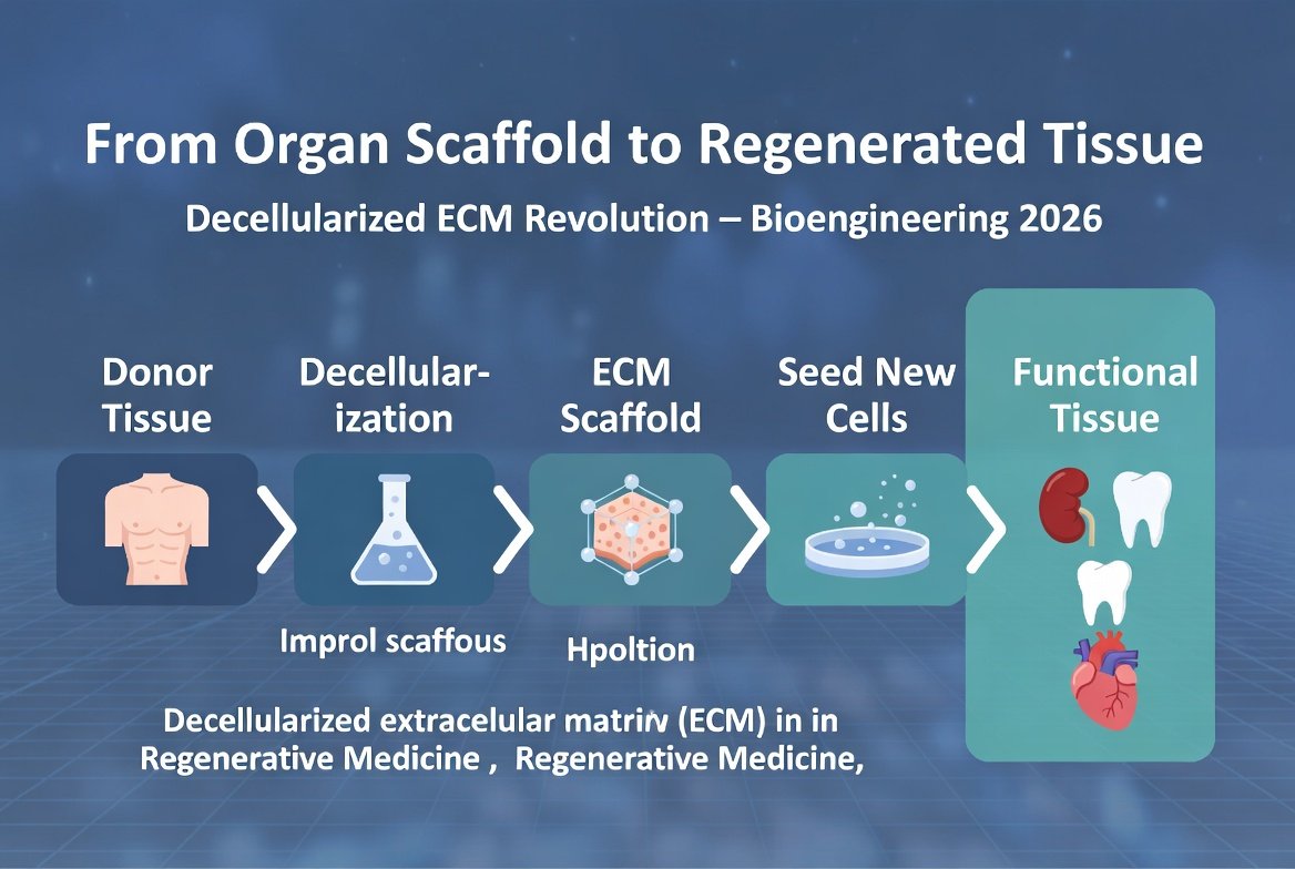
- Extracellular matrix (ECM) scaffold: The three-dimensional structure of an organ, including collagen, elastin, and laminin, without living cells. Imagine a house without tenants.
- Stem cells: Usually iPSCs, induced pluripotent stem cells, reprogrammed from skin or blood cells.
- Bioreactor: A device that simulates physiological conditions, fluid flow, oxygen, and temperature, allowing cells to divide and differentiate within the scaffold.
The theoretical advantage of an organ derived from the patient's own cells is that immunologically it is part of the patient, which could reduce the need for lifelong immunosuppressive drugs and the risk of rejection. But it is important to emphasize: engineering a whole, functional organ from stem cells has not yet been achieved in humans, and it is a tremendous scientific challenge.
The connection to transplant medicine: bridging the gap
To understand why these technologies are important, one must understand the gap between the two worlds.
Classic transplant medicine is built on transferring a living organ from one person to another. It works, it saves lives, but it depends on donors. The demand for organs is growing faster than supply, mainly because the population is aging. The average wait for a kidney in the US often lasts several years.
Regenerative medicine tries to break this barrier from two directions. The first direction, already practical today, is expanding the donor pool by repairing organs through perfusion, so that organs that would have been rejected in the past become usable. This is exactly what the Canadian lab and the EVLP system do. The second direction, more distant, is tissue and organ engineering from stem cells, which is still mainly in the basic research stage.
Decellularization: taking an organ and breaking down only the cells
One of the techniques being researched in tissue engineering is decellularization, first developed by Doris Taylor and her team in 2008. The idea: take an organ and wash it with detergent agents like SDS, which remove all cell membranes and DNA, but leave intact the extracellular matrix scaffold, that three-dimensional network of proteins that makes up the organ's structure.
The result is a transparent-white 'ghost organ', devoid of cells, but with all the original geometry: blood vessels, internal structure, and valves. It is like getting a ready-made house skeleton, full of floors and rooms, just without tenants. The advantage: the natural scaffold preserves the blood vessel network, which is one of the hardest problems in tissue engineering.
Where does the evidence actually stand?
It is important to separate what has been proven from what is still a vision. These are the real, verified findings:
Proof of concept: regenerated rat heart (2008)
This was the foundational proof of concept. Doris Taylor's team decellularized an adult rat heart, repopulated the scaffold with heart cells and endothelial cells, and made it beat again in a bioreactor. By day eight, under load and electrical stimulation, the engineered heart produced a pumping output equal to about 2% of an adult heart (or about 25% of a 16-week fetal heart). Very little, but it was proof that the approach is possible. The study was published in Nature Medicine and became one of the most cited works in the field.
Xenotransplantation: genetically engineered pig kidneys in humans (2024 and 2025)
A completely different technology, not decellularization, has made impressive progress. In March 2024, Massachusetts General Hospital performed the world's first transplant of a CRISPR genetically engineered pig kidney into a living human. In January 2025, a second transplant was performed at this center, in a 66-year-old patient who had been on dialysis for about two years, and he was discharged from the hospital about a week after surgery. In these kidneys, pig genes were inactivated or edited, and human genes were added to improve compatibility and reduce rejection. This is not a kidney grown from the patient's cells, but a genetically engineered pig organ, so it still requires immunosuppression. Nevertheless, it is one of the most significant advances in the field at this time.
Bioprinting kidney tissue: ARPA-H grant (2026)
In January 2026, the Wake Forest Institute for Regenerative Medicine received a grant from the US agency ARPA-H, totaling up to $24.8 million over five years, as part of the PRINT program. The goal: to develop a three-dimensionally printed kidney tissue with blood vessels, which will support kidney function in patients with kidney disease. The tissue is intended to be made from the patient's own cells combined with bio-ink. This is an early stage of research, a tissue that supplements function, not a whole functioning kidney.
What are the real challenges?
The enthusiasm is legitimate, but there are serious caveats worth knowing.
The gap between model and humans
Most studies in whole organ engineering have been done in animals or on isolated tissues. Humans are far more complex, live a long time, and require organs that will function for decades, not days or weeks. An approach that works in the lab does not guarantee a whole, functional organ in a human.
Cancer risk from iPSCs
iPSCs, cells reprogrammed to be pluripotent, carry a theoretical risk. If a cell does not fully differentiate and grows uncontrollably, it could become a teratoma, a tumor containing multiple cell types. This risk is managed through strict quality control, but it cannot be ignored.
Ethics
Some experimental approaches raise deep ethical questions, for example regarding the use of animals or chimeras. Most groups move cautiously in this area under regulatory oversight.
Realistic timeline
Repair and preservation of organs through perfusion are already in the clinic today. Genetically engineered pig kidney transplants are at the stage of individual patients and early clinical trials. Engineering a whole, functional organ from stem cells, on the other hand, is still many years from the clinic, if it ever arrives. One should be wary of headlines promising "organs on demand" soon.
What can be done in the meantime?
- If you are on a transplant waiting list, current treatment, transplantation from a donor, remains the best chance. Regenerative technologies are promising, but most are still far from the clinic. Expanding the donor pool through perfusion is the progress that is already helping today.
- Keep your organs healthy. Kidneys, heart, and liver respond well to a healthy lifestyle: a Mediterranean diet, regular physical activity (the common recommendation is about 150 minutes per week), quality sleep, and avoiding smoking. A healthy lifestyle meaningfully reduces the risk of organ failure, even if an exact percentage cannot be quantified.
- Check your kidney function routinely. A creatinine and GFR test can detect problems early, when there is still time to slow decline.
- If you have early-stage chronic kidney disease, act now. Drugs from the SGLT2 inhibitor family (such as dapagliflozin and empagliflozin, based on the DAPA-CKD and EMPA-KIDNEY studies) and finerenone (based on the FIDELIO-DKD study) have been shown to slow kidney decline. A conversation with a nephrologist is critical.
- Consider donating organs. Today, people die on the waiting list. Marking organ donation or signing a donor card is an act that can save many lives.
- Avoid nephrotoxic drugs if possible. NSAIDs (ibuprofen, naproxen) in high doses and over long periods, and contrast agents in imaging tests, can damage the kidneys, especially if they are already weak. Consult a doctor.
The broader perspective
The story of the organ regeneration lab is not just a story about organs. It marks a gradual shift in how we think about transplant medicine. Instead of settling for only perfect organs, we are learning to repair and rehabilitate damaged organs to expand the pool. At the same time, basic research in tissue engineering is exploring whether one day we can grow tissues, and perhaps in the distant future organs, in the lab.
It is important to keep perspective. These technologies will not replace a healthy diet, physical activity, or quality sleep as the foundations of health. They are an additional tool in the toolbox, not a substitute. A person who keeps their organs healthy reduces from the outset the chance of needing a transplant.
And even if the more ambitious part of the vision, whole organs from the patient's cells, is still far off, the progress in repairing organs through perfusion is already saving lives today. This is a reminder that the great revolutions in medicine are often built step by step, not in one leap.
Organs repaired and preserved outside the body are, therefore, not just a technical novelty. They are a change in the perception of what can be done with a donor organ, and how many people can be saved from that limited pool.
References:
Hospital News - Saving Organs, Saving Lives: Building the World's First Organ Regeneration Lab
Google News - Original Article


💬 Comments (0)
Be the first to comment on the article.